Gluteus Medius Tendon Repair
Hip abductors are a major group of muscles found in the buttocks. They include the gluteus maximus, gluteus medius, gluteus minimus, and tensor fascia lata muscles.
Gluteus medius is situated on the outer surface of the hip. The function of the gluteus medius is to assist with pelvis stability, hip abduction, along with internal and external rotation of the hip. Tears of the gluteus medius usually occur where the tendon inserts at the greater trochanter, causing lateral hip pain.
Tears of the gluteus medius can occur due to traumatic injury or degenerative conditions such as tendinopathy (chronic inflammation of the gluteus medius tendon). Gluteus medius tears cause pain and weakness on the affected side of the hip. One of the main symptoms of a gluteus medius tear is the presence of Trendelenburg sign – dropping of the pelvis towards the unaffected side by being unable to bear weight on the affected limb.
The diagnosis of gluteus medius tear is based on physical examination of the patient, followed by palpation of the affected muscle, testing muscle power and assessing walking pattern or gait of the patient. Certain special tests such as single-leg squat test or a positive Trendelenburg sign confirm the diagnosis of gluteus medius tear. Sometimes, MRI or ultrasound may be helpful to show the pathological changes of the muscle.
Treatment
The aim of treatment is to restore the normal function of the gluteus medius tendon.
- Immediately following the rupture of the tendon, you should do the following:
- Rest your hip by refraining from activities until it is healed.
- Apply ice to your hip to reduce pain and inflammation caused by injury.
- Elevation involves keeping the affected hip raised above your heart to minimize swelling.
- Medications such as non-steroidal anti-inflammatory drugs (NSAIDs) or steroid injections may be given to reduce the pain and inflammation.
- Assistive devices such as a cane or crutch may be used temporarily to facilitate pain free ambulation.
- Your surgeon may recommend physical therapy to strengthen the muscles and increase stability of the hip.
- Surgical treatment may be recommended to repair a complete, full-thickness gluteus medius tear. The rupture can be repaired arthroscopically to help restore the strength and function of the gluteus medius.
Gluteus medius tendon ruptures can be repaired by open or arthroscopic technique.
The steps involved in open surgery include:
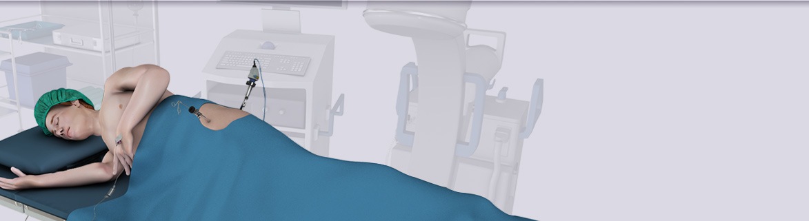
- You will be placed either under general anesthesia or spinal anesthesia
- The surgeon makes a long incision in your hip.
- The underlying muscles and tissues are separated to expose the damaged tendon.
Arthroscopic surgery involves the following steps:
- You will be placed either under general anesthesia or spinal anesthesia
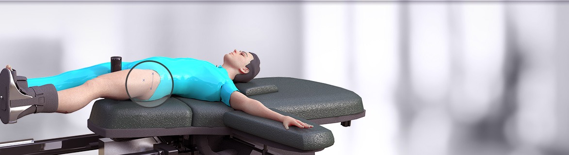
- The surgeon makes two or three small incisions, about 1/4 of an inch each, around the hip joint area. Each incision is called a portal. A blunt tube, called a trocar, is inserted into each portal prior to the insertion of the arthroscope and surgical instruments.
- The arthroscope is inserted through one of the portals, to view the hip joint. The images from the arthroscope guide the surgeon and assist in detection of any anomaly. It also enables the surgeon to see the attachment of the gluteus medius muscle.
- The surgeon conducts a diagnostic endoscopic examination of the peritrochanteric space.
- The other portals are used for the insertion of surgical instruments.
The following steps are common for both open and keyhole surgeries:
- Debridement of the tears on the under surface of the gluteus tendon and pathologic tendon tissue is performed using a shaver.
- The tear at the lateral facet of the greater trochanter is identified and removed using a bur to stimulate active bleeding which would aid in healing of the repaired tendon
- The tendons are visualized and an anchor(s) is/are placed onto the greater trochanter of the femur and a suture is passed through the ruptured tendon. The tendon is then pulled down to its normal anatomic position and tied over the bone.
- After the surgical procedure is complete, the incisions will be closed with sutures or surgical tape.